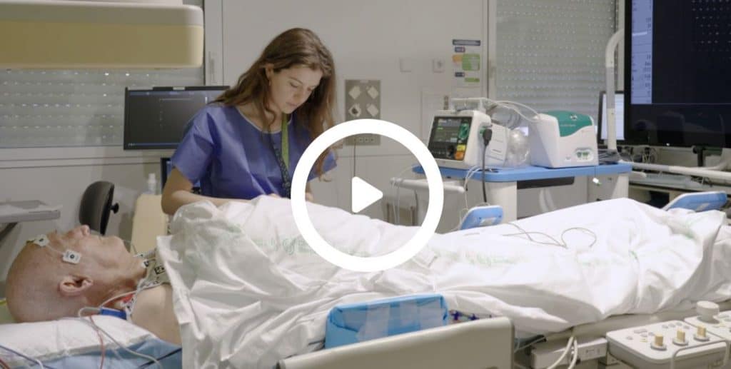
Our work has been featured in the Journal of Visualized Experiments (JoVE) — including a full video that shows, step by step, how Corify’s non-invasive, imageless ECGI system is used in clinical practice.
In this publication, Jana Reventós together with Dr. Lluis Mont (Hospital Clínic), and Dr. Ivo Roca, and members of the Corify team demonstrate how our technology captures electrical activity from the body surface and reconstructs 3D activation maps of the heart, in real time — without CT or MRI. This study has been part of the SAVECOR project funded by EIT HEALTH.
 What You’ll See in the Video
The JoVE article includes a detailed walkthrough of the full mapping process:
- Preparation and electrode placement on the patient
- Data acquisition and system interface
- Real-time generation of 3D electrical activation maps
All using only surface electrodes and our computational modeling platform.
 What Makes Corify Different
- No imaging required: No need for MRI or CT
- Fast setup: Under 10 minutes
- Real-time results: Beat-by-beat activation mapping
- Fully non-invasive: Safe and repeatable, even for follow-up
Designed to complement invasive procedures, our system helps clinicians localize arrhythmia sources faster, with minimal setup and zero radiation exposure.
 A Practical Contribution to Clinical Electrophysiology
We believe medical technology should be intuitive, scalable, and designed for real clinical environments. This video offers a transparent, visual explanation of how ECGI can be applied in practice — something especially valuable for hospitals, researchers and new clinical users.


